Dr Ben Esdaile
Consultant Dermatologist
MBBS (Hons) BSc MRCP
Dermatofibromas:
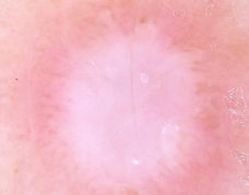
The most common dermoscopic features of dermatofibromas are the central white scar-like area with a faint peripheral network. Polarising dermoscopy may show chrysalis-like structures to the collagen formation centrally. Small clusters of blood vessels are often seen centrally. Ring-like globules may be seen centrally.
Blue naevi:
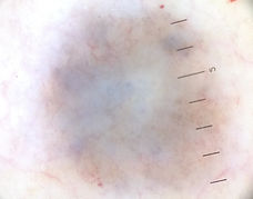
The most common dermoscopic features of blue naevi is a homogenous slate-blue pigment pattern. In the sclerotic variants then a white/blue homogenous pigment is seen. In a combined naevus an eccentric homogenous blue pigment blotch is seen with the rest of the naevus having a benign naevus appearance (combined naevi are great mimickers of melanoma).
There are some other variants of blue naevi such as sclerotic (homogenous white often with chrysallis-like structures on PD), aneurysmal and deep penetrating.
Other benign skin lesions.
A dermatofibroma showing a central scar-like area with faint peripheral network.

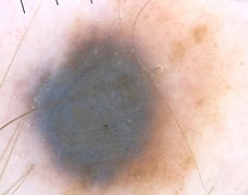
A blue naevus showing homogenous blue pigment with a peripheral brownish hue.
A combined naevus showing an eccentric blue blotch.
A blue naevus showing a heterogenous pigment with blue, white and brown colours.