Dr Ben Esdaile
Consultant Dermatologist
MBBS (Hons) BSc MRCP
Seborrhoeic keratoses and related lesions.
Solar lentigines:
Fingerprint-like structures:
As the name suggests these are parallel lines forming the shape of a fingerprint.
Moth-eaten border:
The edge of the lesion appears to have been eaten by a moth.
Reticular network:
Some lentigos (as seen above) will form a reticular network due to the melanin filled keratinocytes.
Symmetrical follicular pigmentation:
Pigmentation in solar lentigines is symmetrical around hair follicles forming brown circles.
Seborrhoeic keratoses:
Early flat seborrhoeic keratoses have the same dermoscopic features as solar lentigenes.
Typical features of seb k's include:
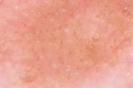
Milia-like cysts:
These are keratin cysts within the lesion that appear as milky white blobs. They can also appear in naevi and basal cell carcinomas.
Hairpin vessels:
These vessels form a 'u' as they bend back on themselves often with a surrounding white halo from the keratin.
Comedo-like openings:
These are essentially keratin plugs in the craters of the surface of the lesion.
Ceribriform stuctures:
The ridges and sulci of the undulating surface of a seborrhoeic keratois gives rise to the 'brain-like' appearance of some seborrhoeic keratoses.
Lichenoid keratoses:
Thought to result from an immune reaction to either solar lentigines, seborrhoeic keratoses or actinic keratoses. Dermoscopy often reveals diffuse coarse or fine granular pigmentation. It can sometimes be hard to differentiate from the fine granular pigmentation of regression.
Symmetrical follicular pigmentation in a solar lentigo.

A Seborrhoeic keratosis showing milia-like cysts, hairpin vessels and comedo-like openings.
The cerebriform structures of a seborrhoiec keratosis


Fingerprint-like structures in a flat lentigo/ evolving seborrhoeic keratosis
Moth-eaten border of a solar lentigo.


Dermoscopy showing diffuse fine granular pigmentation in a lichenoid keratosis.

An irritated seborrhoeic keratosis showing more prominent vascular markings (hairpin vessels).